The Role of Radiography in Dental Diagnosis
11/03/2026 Share :
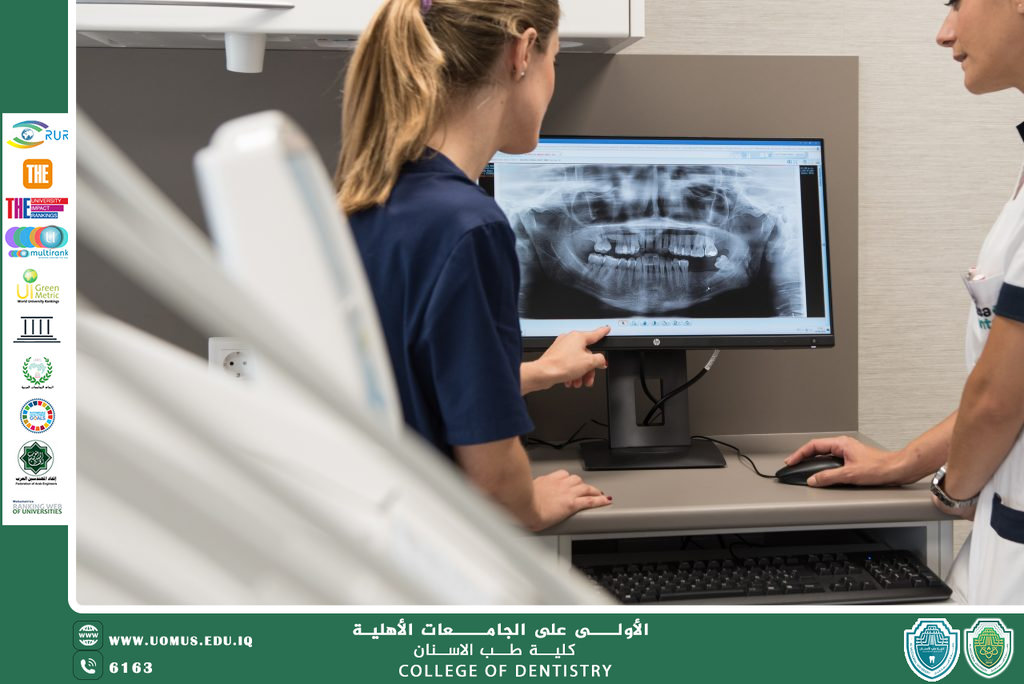
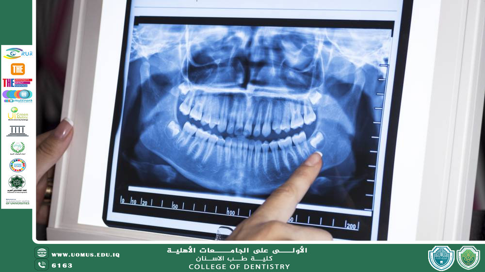
Accurate diagnosis is the first step toward successful dental treatment, and in many cases it cannot be achieved through clinical examination alone. Dental radiography therefore plays a crucial role by allowing dentists to visualize structures that are not visible to the naked eye.
Dental X-rays enable the detection of many hidden conditions within teeth and surrounding bone structures. They help identify deep dental caries, evaluate the health of the supporting bone, diagnose root infections, and determine the position of impacted teeth or developmental dental abnormalities.
Radiographic imaging also plays a key role in treatment planning. Whether in endodontic therapy, oral surgery, orthodontics, or dental implant procedures, radiographs provide dentists with a clear understanding of the anatomical structures involved. This information helps clinicians choose the most appropriate treatment approach and avoid potential complications.
In recent years, dental imaging technologies have advanced significantly. Digital radiography now provides clearer images while reducing radiation exposure to patients. These systems also allow images to be stored, analyzed, and compared over time, which is particularly useful for monitoring disease progression and treatment outcomes.
Despite its importance, radiographic examination must always follow the principle of balancing diagnostic benefit with patient safety. Dentists request radiographs only when clinically justified and apply protective measures to minimize radiation exposure.
Ultimately, dental radiography has become an indispensable diagnostic tool in modern dentistry. It enables early detection of oral diseases and supports precise treatment decisions, thereby improving the overall quality of dental care.