The Role of Ultrasound Elastography in the Diagnosis of Uterine Masses among Iraqi Women
31/03/2026 Share :
Asst. Lecturer Ahmed Salman Jassim
Department of Radiological Techniques, College of Health and Medical Technologies, Al-Mustaqbal University, Babylon, Iraq
Uterine masses are among the most common gynecological conditions affecting women of reproductive age. These include uterine fibroids, adenomyosis, endometrial polyps, and malignant tumors. Ultrasound imaging is widely used as a primary diagnostic modality due to its availability, non-invasive nature, and relatively low cost. However, conventional ultrasound may sometimes fail to clearly differentiate between various types of uterine lesions. Ultrasound elastography is an advanced imaging technique that measures tissue stiffness and elasticity, providing additional diagnostic information. This paper reviews the clinical role of ultrasound elastography in the diagnosis of uterine masses.
Keywords:
Ultrasound elastography, uterine masses, fibroids, adenomyosis, diagnostic imaging, women’s health
1. Introduction
Uterine masses represent one of the most common gynecological findings among women worldwide. The prevalence of uterine fibroids is estimated to affect between 20% and 50% of women during their reproductive years. Other conditions such as adenomyosis, endometrial polyps, and malignancies may also present as uterine masses. Early diagnosis is essential for appropriate clinical management and prevention of complications such as infertility, abnormal uterine bleeding, and pelvic pain.
2. Principles of Ultrasound Elastography
Ultrasound elastography is a technique used to assess the mechanical properties of biological tissues. It is based on the principle that different tissues exhibit different elastic properties; softer tissues deform more easily than harder tissues when mechanical pressure is applied. The degree of deformation is then converted into a color-coded image representing tissue stiffness.
3. Ultrasound Elastography in the Evaluation of Uterine Masses
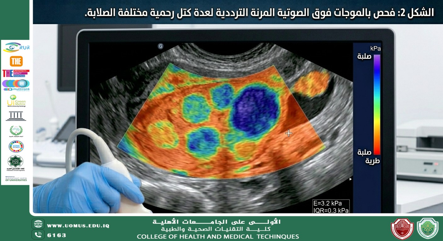
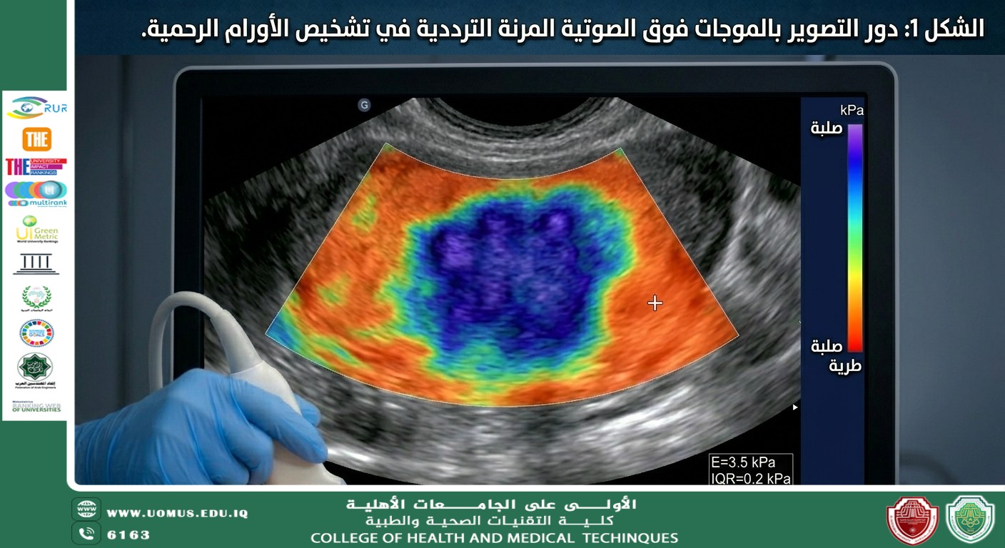
Different uterine pathologies exhibit varying stiffness patterns when assessed باستخدام ultrasound elastography. Uterine fibroids are typically stiffer than normal myometrium, while adenomyosis appears as heterogeneous areas with intermediate stiffness.
5. Clinical Significance in Iraq
In Iraq, ultrasound is widely used in public hospitals and private centers. Access to advanced imaging modalities such as MRI may be limited due to high costs. Ultrasound elastography provides a practical solution to enhance diagnostic accuracy without the need for expensive equipment.
6. Advantages and Limitations
The advantages of ultrasound elastography include improved lesion characterization, non-invasive assessment, real-time imaging, and relatively low cost. However, the results may depend on operator experience and patient-related factors.