
Scientific article dr.emad entitled Ameloblastoma
16/11/2019 Share :
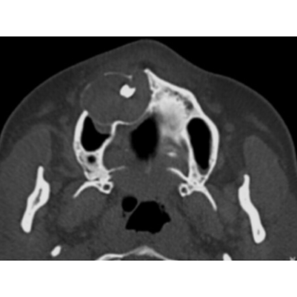
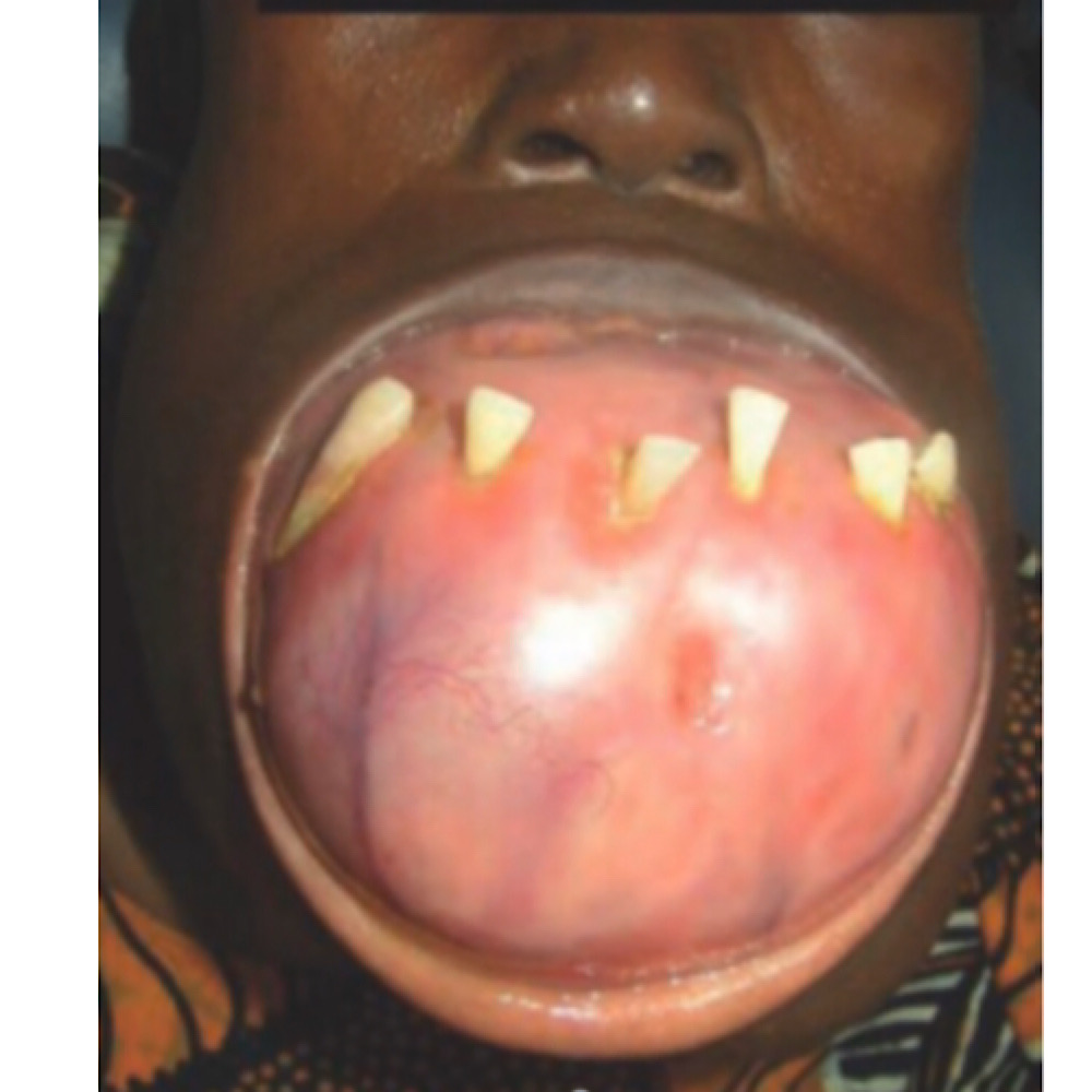
<br />The ameloblastoma is the most common clinically significant and potentially lethal odontogenic tumor. Excluding odontomas, its incidence equals or exceeds the combined total of all other odontogenic tumors. May arise from:<br /> 1. Rests of the dental lamina,<br />2. A developing enamel organ,<br />3. The epithelial lining of an odontogenic cyst,<br />4. The basal cells of the oral mucosa.<br /> Three different variants, each with specific implications for treatment and a unique prognosis: solid or multicystic, unicystic, and peripheral. The solid and multicystic forms of the tumor do not produce an effective connective tissue capsule or evoke an encapsulating mechanism by surrounding bone or soft tissue The unicystic ameloblastoma, however, does have a peripheral connective tissue wall, which may be loose or dense, and relatively thin or thick<br />Treatment:<br />The treatment is surgical resection with a safety margin. A safe margin of uninvolved bone is<br /> approximately 1.5 to 2 cm for solid and multicystic lesions, and 1 to 1.5 cm for unicystic and peripheral lesions. Post-treatment patients should be followed for 15 to 20 years or longer due to high recurrence rate and slowly growing behavior of tumor.<br /> Immediate reconstruction can be performed if there is surgical, specimen radiographic, or intraoperative frozen section certainty of complete excision of the tumor. However, if there is uncertainty about the resection margins, reconstruction should be delayed until permanent tissue sections are studied. Maxillary lesions must be resected aggressively because of the ease with which the tumor can spread through the less dense cancellous and cortical bone, with access to the pterygoid region, sinuses, infratemporal space, and skull base.<br /> <br /> Although some studies suggest that the ameloblastoma may be radiosensitive, radiation therapy has seldom been used as a treatment modality because of the intraosseous location of the tumor and the potential for secondary radiation-induced malignancy developing in a relatively young patient population.