
An article about rickets by lecturer Elaf Ali Obaid
19/03/2024 Share :
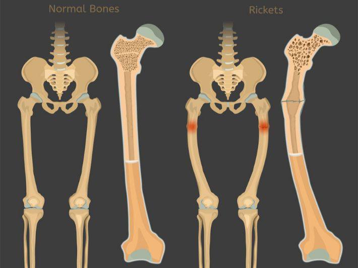
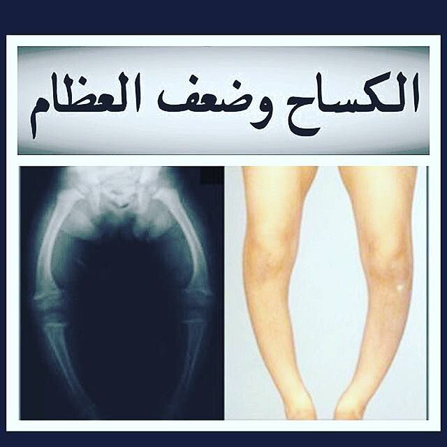
Baby rickets, or reck, is a condition that leads to weakness or loose bones in children, caused by either nutritional deficiencies or genetic causes that include curvature of the legs, growth distress, bone pain, large forehead, and difficulties in sleep. Complications may include bone deformities, false bone fractures, abnormally curved muscle or spine cramps<br /><br />The most common cause of rickets is vitamin D deficiency, although there are also genetic forms. This can result from eating a diet that does not contain enough vitamin D, dark skin, lack of sun exposure, exclusive breastfeeding vitamin D supplements, celiacosis, and some genetic conditions. Other factors may include insufficient calcium or phosphorus including the underlying mechanism insufficient calcification of the growth plate. The diagnosis is generally based on blood tests showing low calcium, low phosphorus, and high alkaline phosphatase with X-rays<br /><br />Prevention for children who are exclusively breastfed are vitamin D supplements. Otherwise, treatment depends on the underlying cause if it is due to a lack of vitamin D, treatment with vitamin D and calcium is usually this generally leads to improvements within a few weeks. Bone deformities may also improve over time from time to time Surgery to correct bone deformities The genetic forms of the disease usually require specialized treatment<br /><br />Rickets occur relatively commonly in the Middle East, Africa and Asia. It is generally uncommon in the United States and Europe, with the exception of some minority groups starting in childhood, usually ranging from 3 to 18 months of age with equal disease rates between males and females. Cases of what is believed to be a ricket have been described since the first century, the condition was widespread in the Roman Empire Early treatments included the use of cod liver oil<br />The most important and common causes of this disease were found, the most important and common of which is the lack of vitamin D because the most important function of this vitamin is to increase the levels of calcium and phosphorus in the blood by increasing the absorption of calcium and phosphorus salts from the intestine and reducing their secretion with the urine, and then their transmission to build bones and convert the soft cartilage parts of them into solid bone parts, which allows the construction of the skeleton. Because the gland hormone of the thyroid hormone helps to synthesize vitamin D, the lack of activity of this hormone is a major cause of a lack of calcium salts and bone play.<br /><br />There are other causes of bone lenosis other than dysfunction or synthesis of vitamin D function, including chronic liver or kidney disease, blood problems, chronic diarrhea, dysfunction of absorption from the small intestine, and the use of some drugs for long periods such as some drugs used to treat cases of epilepsy.<br /><br />Bone soft disease can also be caused by genetic diseases such as those that affecting the kidneys, the activity of its enzymes necessary to make vitamin D or weaken their ability to save phosphate salts in the body.<br /><br />The prevalence of Leen Bone disease increases in the first and second years of the child's age. Symptoms appear after vitamin D deficiency for several months. The severity of the symptoms of the disease increases with the delay of the treatment of the condition or according to its accompanyment to other pathogens, and the most important symptoms of the disease are as follows:<br /><br />The head<br /><br />Looseness: In the areas adjacent to the joints of the skull and the continued expansion of the fontage area with the increase in the size of the head, the protrusion of the forehead and the change of its circular shape, the delay or absence of the appearance or deformation of the teeth<br /><br />The chest<br /><br />• The appearance of rosary-shaped bumps in the ends of the ribs when the cartilage is contacted with the bones.<br /><br />• The protrusion of the breast bones forward to give a pigeon-like shape, which is called the pitral chest.<br /><br />• The presence of a diaphrosis in the lower part of the chest along the attachment of the diaphragm to the chest wall from the inside [8] called a harrison groove[9] and Harrison is the British doctor who described this groove.<br /><br />The spine<br /><br />The spine may experience abnormal lateral or anterior curvatures.<br /><br />The growth of pelvic bones is delayed with various abnormalities.<br /><br />The parties<br /><br />The ends of the limb bones around the wrist and ankle swell with curvatures in the long bones of the upper and lower extremities that appear more clearly in the curvature of the stems or contact of the knees and these deformities in the spine and lower limbs may lead to short stature.<br /><br />Ligaments<br /><br />Joint ligaments experience relaxation and softness.<br /><br />Muscles<br /><br />This disease leads to delayed muscle growth, general weakness and delayed muscle growth in the child so that the child is delayed in crawling, loving, sitting, standing and walking. A lack of calcium salts also leads to muscle contractions and frequent cramps.<br />