A scientific article by the teaching assistant (Hanin Hani) entitled “Behind the image: What happens inside the MRI machine?”
18/05/2025 Share :
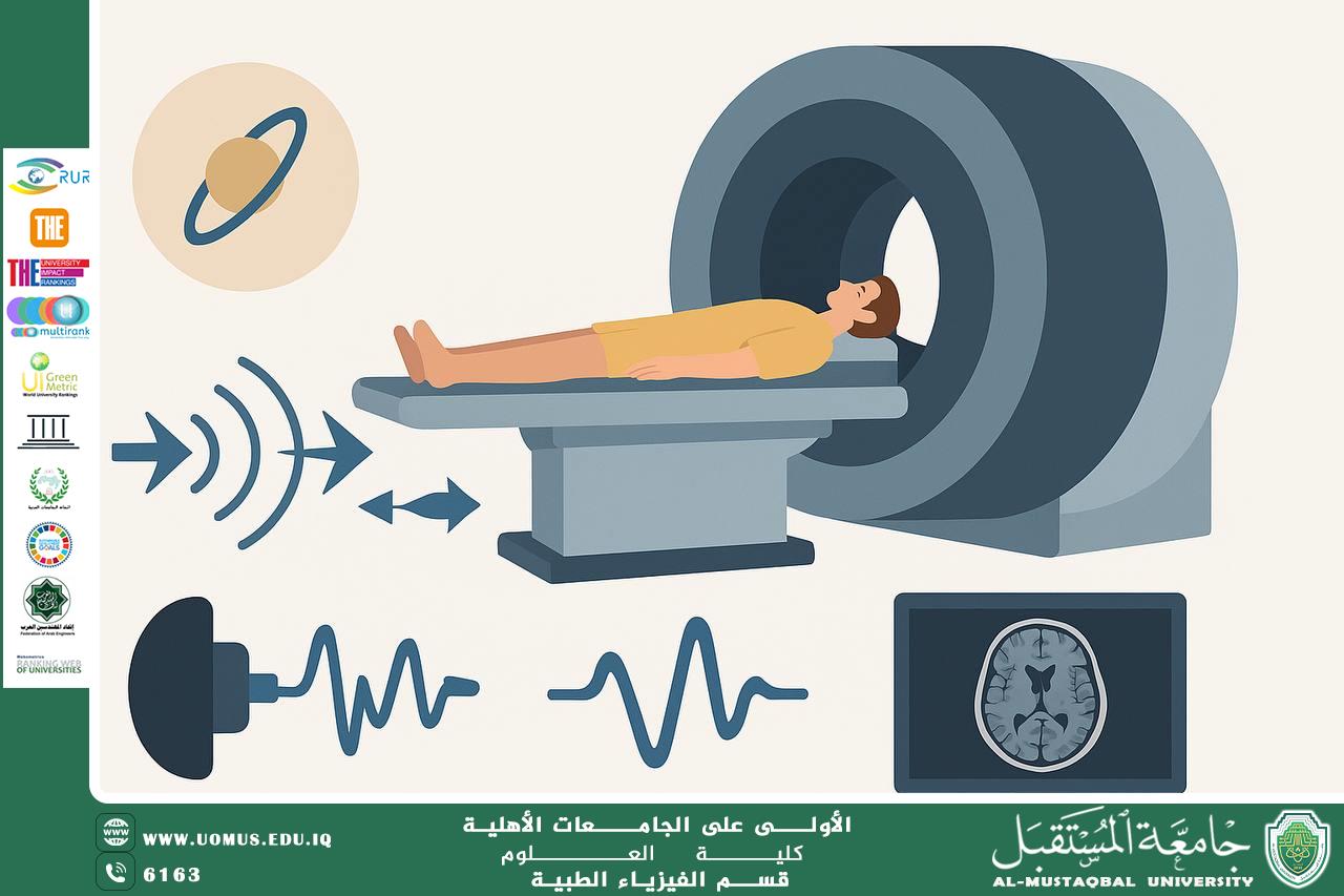
Abstract:<br /><br />Magnetic Resonance Imaging (MRI) is one of the most powerful tools in medical diagnostics, providing detailed images of internal body structures without the need for harmful radiation. While the final images are familiar to many, the physics that takes place inside the MRI machine is often a mystery. This article unveils the hidden science behind MRI technology, explaining its core principles in a simple yet scientifically accurate manner, tailored for students and healthcare professionals interested in medical physics.<br /><br />⸻<br /><br />Introduction:<br /><br />At first glance, an MRI scanner looks like a giant doughnut with a sliding bed. But once the patient lies down and the machine hums into action, a beautifully detailed image of the inside of the body appears—organs, tissues, even brain activity. What makes this magical image possible? The answer lies in the fascinating world of nuclear magnetic resonance, a principle rooted in quantum physics and electromagnetic theory.<br /><br />⸻<br /><br />How Does MRI Work?<br /><br />1. The Body and Hydrogen Atoms:<br /><br />The human body is made mostly of water, and water contains hydrogen atoms. These atoms have tiny nuclei (protons) that behave like spinning magnets.<br /><br />2. The Magnetic Field:<br /><br />The MRI scanner contains a powerful magnet—often 1.5 to 3 Tesla, thousands of times stronger than Earth’s magnetic field. When you lie in the scanner, this field aligns the hydrogen protons in your body in the same direction, like compass needles.<br /><br />3. Radiofrequency Pulse:<br /><br />Next, the machine sends a radiofrequency (RF) pulse which “knocks” these aligned protons out of their position. When the pulse stops, the protons relax and realign with the magnetic field, releasing energy as they do.<br /><br />4. Signal Detection and Image Creation:<br /><br />The released energy is detected by the machine and translated into digital signals. A computer processes these signals to create cross-sectional images of the body.<br /><br />Each tissue—fat, muscle, brain, bone—has a unique signal, allowing MRI to differentiate between them with high precision.<br /><br />⸻<br /><br />Why is MRI So Important?<br /> • No Ionizing Radiation: Unlike X-rays or CT scans, MRI is safe for repeated use.<br /> • Soft Tissue Clarity: MRI offers unmatched detail when imaging the brain, spinal cord, joints, and soft tissues.<br /> • Functional Imaging: Advanced MRI techniques (like fMRI) can track blood flow and brain activity in real time.<br /><br />⸻<br /><br />The Sounds and the Experience:<br /><br />Many patients report loud knocking or buzzing sounds during the scan. These come from gradient coils, which rapidly change the magnetic field to locate signals precisely in 3D space.<br /><br />To improve comfort, earplugs and music are often provided. Some scanners now feature ambient lighting or video projection for relaxation.<br /><br />⸻<br /><br />Limitations and Precautions:<br /> • Metal Implants: Pacemakers or metal fragments in the body can pose risks.<br /> • Claustrophobia: The enclosed space can cause discomfort for some.<br /> • High Cost: MRI machines are expensive and require trained staff.<br /><br />⸻<br /><br />Conclusion:<br /><br />MRI is more than just a medical imaging tool—it’s a masterpiece of modern physics and engineering. What happens inside the scanner is an elegant dance of atoms, fields, and signals working together to reveal the hidden beauty of the human body. Understanding the science behind it not only enriches our appreciation but also highlights the role of medical physics in everyday healthcare.<br /><br /><br /><br />"AL_mustaqbal University is the first university in Iraq"<br/><br/><a href=https://uomus.edu.iq/Default.aspx target=_blank>al-mustaqbal University Website</a>