A scientific article by the teaching assistant (Zahraa Bassem) entitled “Physics Under the Skin: How to Read the Body Without Touching It”
18/05/2025 Share :
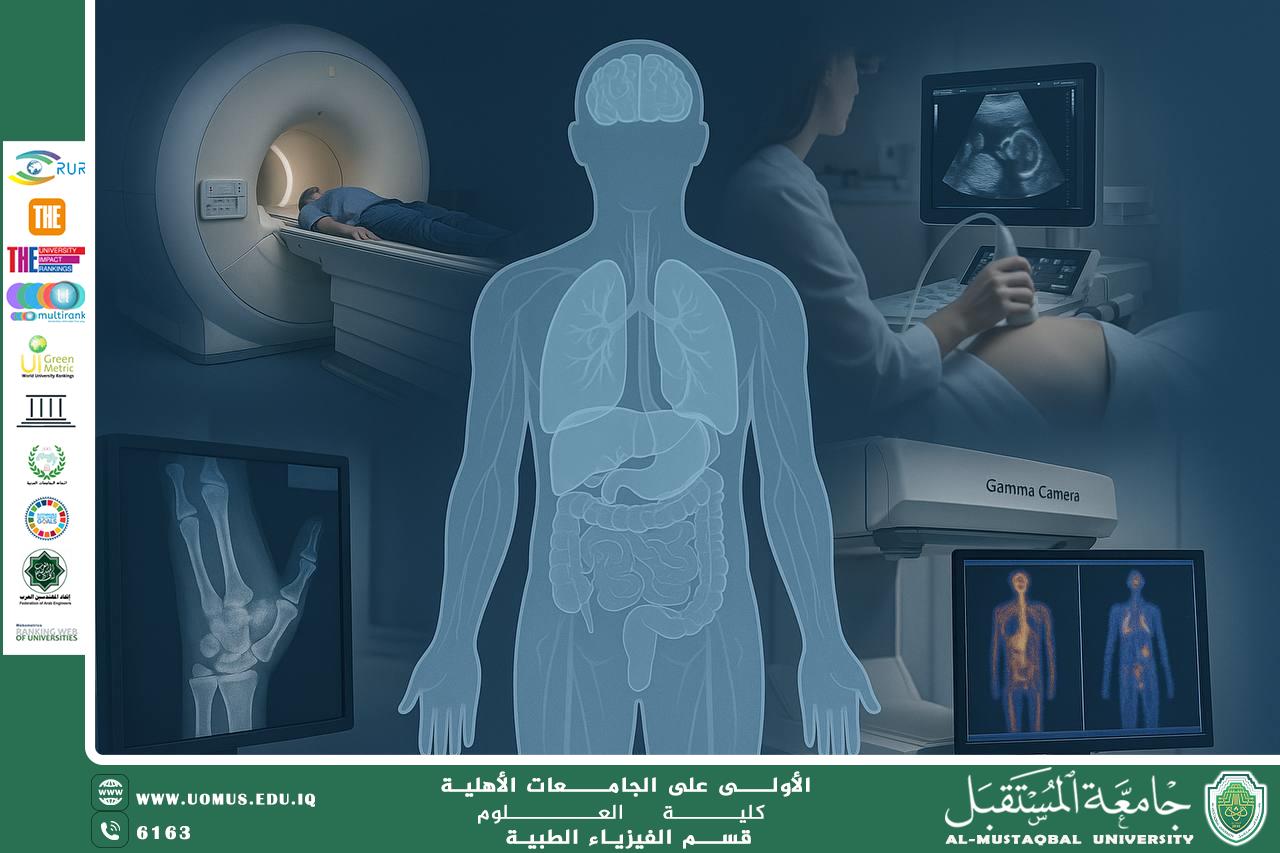
Abstract:<br /><br />In recent decades, the field of medical diagnostics has undergone a major transformation due to the advancement of non-invasive imaging technologies. These innovations—grounded in medical physics—have enabled clinicians to visualize internal organs and tissues with high precision, without the need for surgical intervention. This article provides an academic overview of non-invasive imaging modalities, exploring the underlying physical principles, mechanisms of action, and clinical applications. The objective is to highlight the crucial role of physics in facilitating safe, accurate, and non-invasive diagnostic methods.<br /><br />⸻<br /><br />Introduction:<br /><br />The ability to explore the inner workings of the human body without surgical procedures has long been a core ambition in medicine. Thanks to advances in non-invasive imaging, physicians can now observe physiological processes and detect pathologies without physically breaching the skin. These diagnostic capabilities are underpinned by the science of medical physics, which applies core physical principles—such as electromagnetic and acoustic wave behavior, energy absorption, and nuclear decay—to the development of medical imaging technologies.<br /><br />⸻<br /><br />1. Magnetic Resonance Imaging (MRI): Precision Through Magnetism<br /><br />MRI utilizes the magnetic properties of hydrogen nuclei in water molecules. The patient is placed in a strong magnetic field (typically 1.5 to 3 Tesla), aligning the hydrogen nuclei. Radiofrequency pulses then perturb this alignment, and the emitted signals as the nuclei return to equilibrium are used to construct detailed images.<br /> • Advantages: Excellent contrast for soft tissues; no ionizing radiation; functional imaging capability (fMRI).<br /> • Applications: Brain tumors, spinal cord injuries, musculoskeletal disorders, multiple sclerosis.<br /><br />⸻<br /><br />2. X-ray Imaging: Seeing the Bones with Light<br /><br />X-rays rely on the differential absorption of ionizing electromagnetic radiation by tissues of varying density. The resulting attenuation pattern is captured on a detector to produce images that primarily visualize bone and certain dense structures.<br /> • Advantages: Rapid and widely accessible; useful for skeletal imaging.<br /> • Limitations: Exposure to ionizing radiation; limited contrast for soft tissues.<br /><br />⸻<br /><br />3. Ultrasound Imaging: Listening to the Echoes of the Body<br /><br />This technique emits high-frequency sound waves into the body, which reflect off tissues at different rates depending on their density and composition. The returning echoes are processed to generate real-time images.<br /> • Advantages: Completely safe; portable; real-time imaging; no radiation.<br /> • Applications: Pregnancy monitoring, cardiac assessment, abdominal organs, vascular structures.<br /><br />⸻<br /><br />4. Computed Tomography (CT): Cross-sectional Clarity<br /><br />CT scanning integrates X-ray technology with computer processing to generate cross-sectional images of the body. A rotating X-ray source captures multiple angles, which are reconstructed into a 3D image.<br /> • Advantages: Excellent for internal bleeding, trauma assessment, and detailed bone imaging.<br /> • Limitations: Higher radiation dose compared to standard X-rays; not ideal for all patients.<br /><br />⸻<br /><br />5. Nuclear Medicine Imaging: Beyond Structure, Into Function<br /><br />Techniques such as Positron Emission Tomography (PET) and Gamma Scanning involve the use of radiopharmaceuticals that emit gamma rays. These compounds target specific tissues or metabolic processes, enabling functional imaging rather than merely structural.<br /> • Advantages: Functional assessment of organs; early detection of metabolic disorders and cancers.<br /> • Limitations: Radioactive materials require special handling; expensive and less widely available.<br /><br />⸻<br /><br />Underlying Physical Principles:<br /><br />All non-invasive imaging techniques rely on the interaction of physical forces or particles with biological tissues. Core principles include:<br /> • Propagation and attenuation of electromagnetic or sound waves<br /> • Tissue contrast based on density, atomic number, or molecular behavior<br /> • Digital image processing and signal reconstruction algorithms<br />The application of these principles allows for the generation of diagnostic images with high clarity and specificity, without physical contact or surgical procedures.<br /><br />⸻<br /><br />Conclusion:<br /><br />Medical imaging technologies represent a triumph of physics in service of medicine. Through sophisticated applications of electromagnetic theory, nuclear decay, and wave mechanics, we can “see” beneath the skin—diagnosing, tracking, and understanding disease in a non-invasive, patient-friendly manner. The future of medical diagnostics is likely to be shaped by further integration of physics with artificial intelligence and molecular imaging, promising even greater precision and personalization in patient care.<br /><br /><br /><br /><br />"AL_mustaqbal University is the first university in Iraq"<br/><br/><a href=https://uomus.edu.iq/Default.aspx target=_blank>al-mustaqbal University Website</a>