A scientific article by the lecturer, : Dr. Ghaith Ali Mahmoud (Keratoconus)
22/07/2025 Share :
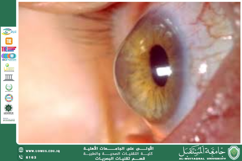
Part Two: Diagnosis and Management<br />Diagnostic Methods<br />The diagnosis of keratoconus relies on a combination of specialized eye examinations, including:<br />• Corneal topography: the most important tool for detecting the cone-shaped distortion of the cornea<br />• Pachymetry: measurement of corneal thickness<br />• Slit-lamp examination: to observe structural abnormalities in the cornea<br />• Refraction and visual quality testing: to assess the degree of visual distortion<br />Disease Staging<br />Keratoconus can be classified into several stages based on severity:<br />• Mild: Slight visual distortion; corrected with glasses or soft contact lenses<br />• Moderate: Requires rigid gas-permeable (RGP) or hybrid lenses<br />• Advanced: Significant loss of visual acuity; may require surgical intervention<br />• Severe: May necessitate full-thickness corneal transplantation<br />Treatment Options<br />Treatment strategy depends on the stage at diagnosis:<br />• Early stages: Managed with eyeglasses or soft contact lenses<br />• Rigid or hybrid contact lenses: Compensate for the irregular corneal surface<br />• Corneal Cross-Linking (CXL): A modern technique that strengthens collagen fibers using ultraviolet (UV) light and riboflavin, helping halt disease progression<br />• Intrastromal Corneal Ring Segments (ICRS): Implanted to reshape and flatten the cornea<br />• Corneal Transplantation: Considered the last resort, involving either partial (DALK) or full-thickness (PKP) grafting depending on the case<br /><br /><br /><br />Recent Advances<br />Recent years have seen remarkable progress in keratoconus treatment technologies, including:<br />• Enhanced CXL techniques, such as epi-on (epithelium-on) protocols that eliminate the need for epithelial removal<br />• Scleral and hybrid lenses, which offer superior visual clarity and comfort<br />• Artificial intelligence algorithms that analyze disease progression and predict future stages<br />• Clinical trials exploring gene therapy and biological treatments aimed at corneal regeneration and tissue healing stimulation<br />