White Sponge Nevus
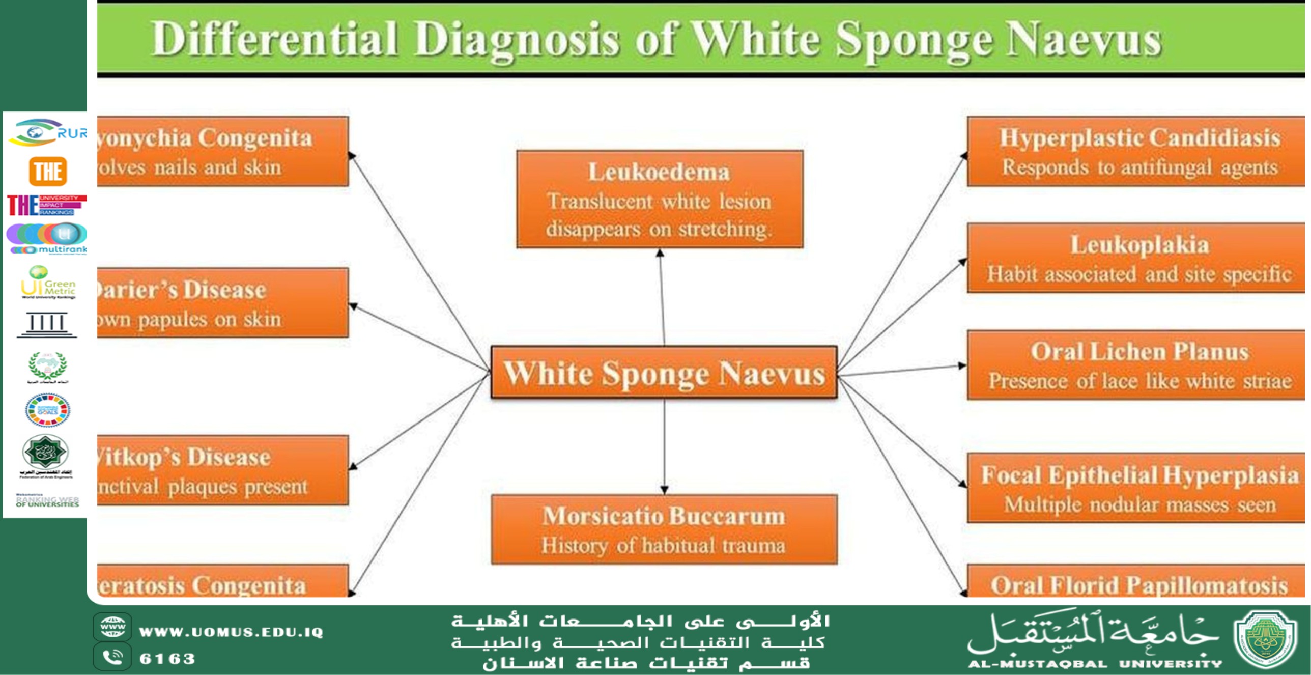
تحقيقا للهدف الثالث وهو الصحة الجيدة والرفاه قام قسم تقنيات صناعة الاسنان بنشر مقاله علمية بعنوان White Sponge Nevus لرئيس القسم أ.د. منى صالح مرزة وذلك يوم الاثنين الموافق 7/4/2025 وتضمنت المقالة:<br />White Sponge Nevus<br />White sponge nevus (WSN) is a rare, benign hereditary condition characterized by thick, white, spongy plaques on the oral mucosa. It is caused by a genetic mutation that affects keratin production, leading to structural abnormalities in the epithelial cells. WSN is a non-malignant condition that typically presents early in life and remains asymptomatic, requiring no specific treatment.<br />Etiology and Pathogenesis<br />White sponge nevus is inherited in an autosomal dominant manner, with mutations in the keratin 4 (KRT4) or keratin 13 (KRT13) genes. These genes are responsible for producing keratins that maintain the structural integrity of epithelial cells in non-keratinized mucosa. Mutations in these genes result in the defective keratin network, leading to epithelial thickening and the characteristic spongy appearance of WSN.<br />________________________________________<br />Clinical Features<br />The clinical presentation of WSN includes:<br />• Appearance: Thick, white, folded or spongy plaques with a velvety or corrugated texture.<br />• Location: Most commonly found on the buccal mucosa (inner cheeks) but may also affect the labial mucosa, tongue, floor of the mouth, and other mucosal surfaces.<br />• Bilateral Symmetry: The lesions are typically symmetrical.<br />• Onset: WSN usually manifests in childhood or early adolescence, often during infancy.<br />• Symptoms: The condition is generally asymptomatic, though some individuals may report mild irritation or sensitivity.<br />Unlike other oral white lesions, WSN is persistent and does not disappear upon stretching the mucosa.<br />It is essential to distinguish WSN from other oral white lesions, including:<br />• Leukoplakia: A premalignant lesion with persistent white patches that may show dysplasia on biopsy.<br />• Oral Candidiasis: White patches that can be wiped off, often associated with erythema or burning.<br />• Lichen Planus: Presents as white, lace-like patterns (Wickham striae) and may involve erosive or ulcerative lesions.<br />• Leukoedema: A diffuse, grayish-white mucosal change that disappears upon stretching.<br />________________________________________<br /><br />The diagnosis of WSN is primarily clinical, based on its characteristic appearance and familial history. A biopsy may be performed in uncertain cases to confirm the diagnosis, revealing histopathological features such as:<br />• Thickened epithelium with parakeratosis and acanthosis.<br />• Intracellular edema within the epithelium.<br />• Eosinophilic perinuclear condensation in epithelial cells.<br />Genetic testing can identify mutations in KRT4 or KRT13 but is rarely needed for routine diagnosis.<br />________________________________________<br />Management<br />WSN is a benign and asymptomatic condition that does not require treatment. However, if irritation occurs or if secondary infections are suspected, symptomatic management may include:<br />• Good oral hygiene to prevent superimposed infections.<br />• Antifungal agents if secondary candidiasis is present.<br />Patient education is crucial to alleviate concerns about the condition’s benign nature.<br />________________________________________<br />Prognosis<br />White sponge nevus is a lifelong condition with an excellent prognosis. It does not increase the risk of malignancy and typically does not impact the individual’s quality of life.<br />________________________________________<br />Conclusion<br />White sponge nevus is a rare genetic condition that manifests as white, spongy plaques in the oral cavity. Recognizing its clinical and histopathological features is essential for distinguishing it from other white lesions. As it is benign and asymptomatic, treatment is rarely required, and patient reassurance remains the cornerstone of management.<br />Prof.Dr. Muna S Merza<br /><br />جامعة المستقبل الجامعة الاولى في العراق