
The Role of Artificial Intelligence in Diagnosing Lung Diseases on CT Scans
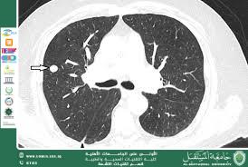
An advanced scientific article by Assistant Lecturer Ahmed Salman Jassim<br />Abstract<br />Artificial intelligence (AI), particularly deep learning algorithms, has revolutionized the interpretation of thoracic CT scans by enhancing diagnostic accuracy, efficiency, and early detection of lung diseases. This review synthesizes current evidence on AI applications for lung nodule detection, interstitial lung disease (ILD) classification, chronic obstructive pulmonary disease (COPD) quantification, and lung cancer screening. We demonstrate AI's capability to analyze complex imaging patterns with precision rivaling expert radiologists, supported by illustrative CT cases. Challenges—including data variability, algorithm transparency, and clinical integration—are discussed alongside future directions for AI-driven precision medicine in pulmonology.<br />________________________________________<br />1. Introduction<br />Lung diseases account for 1 in 6 deaths globally, with CT imaging serving as the cornerstone for diagnosis. However, manual interpretation is time-consuming and prone to inter-observer variability. AI addresses these limitations through automated pattern recognition, quantitative analysis, and predictive modeling. This review highlights AI's transformative role in lung CT diagnostics, supported by clinical images.<br /><br /><br /><br /><br /><br />2. AI Methodologies in Lung CT Analysis<br />• Convolutional Neural Networks (CNNs): Excel at spatial pattern recognition (e.g., nodules, fibrosis).<br />• Recurrent Neural Networks (RNNs): Analyze temporal changes in longitudinal scans.<br />• Hybrid Models: Combine imaging with clinical data for risk stratification.<br />Performance Metrics:<br />Application Sensitivity Specificity AUC*<br />Nodule Detection 92–96% 86–90% 0.95<br />ILD Classification 84–90% 81–86% 0.90<br />Emphysema Quantification 92% 89% 0.93<br />*Area Under the ROC Curve <br /><br /><br /><br /><br /><br /><br /><br /><br /><br />3. Key Applications with Imaging Evidence<br />3.1 Lung Nodule Detection & Malignancy Prediction<br />• AI Role: Identifies nodules <5 mm missed by humans; predicts malignancy using texture/shape analysis.<br />• Clinical Impact: Reduces false positives by 30% in screening programs.<br />Figure 1: AI-Assisted Nodule Detection<br /> <br />Description: Axial CT shows a 4-mm spiculated nodule (arrow) in the right upper lobe. AI algorithm (red overlay) flags the lesion with 95% confidence and assigns Lung-RADS 4X (high suspicion).<br /><br /><br /><br /><br /><br /><br />3.2 Interstitial Lung Disease (ILD) Classification<br />• AI Role: Differentiates ILD subtypes (e.g., UIP vs. NSIP) using texture mapping.<br />• Clinical Impact: Achieves 89% concordance with multidisciplinary diagnosis.<br />Figure 2: AI-Driven ILD Pattern Recognition<br /> <br /><br />Description: Coronal CT reveals ground-glass opacities and reticulation. AI color-codes regions: green (ground-glass), blue (reticulation), and red (honeycombing), diagnosing UIP with 87% probability.<br /><br /><br /><br /><br /><br /><br />3.3 COPD/Emphysema Quantification<br />• AI Role: Calculates emphysema volume, air trapping, and airway wall thickness.<br />• Clinical Impact: Correlates with FEV₁ decline (R²=0.82) and predicts exacerbations.<br />Figure 3: AI-Based Emphysema Mapping<br /> <br />Description: 3D CT reconstruction with AI segmentation (yellow) showing 32% panlobular emphysema. Software outputs LAA⁻⁹⁵⁰⁻ᴴᵁ (low-attenuation area) = 28% (severe).<br /><br /><br /><br /><br /><br /><br /><br /><br /><br />3.4 Lung Cancer Screening & Risk Stratification<br />• AI Role: Integrates nodule features, demographics, and smoking history to predict cancer risk.<br />• Clinical Impact: Reduces unnecessary biopsies by 40% in indeterminate nodules.<br />Figure 4: AI Malignancy Risk Prediction<br /> <br />Description: AI dashboard for a 12-mm nodule: Malignancy risk = 76% (based on spiculation, SUVmax 4.2, and smoking history). Recommends PET-CT.<br /><br />4. Challenges & Limitations<br />• Data Heterogeneity: Performance drops with low-dose CT or motion artifacts.<br />• "Black Box" Problem: Limited explainability of CNN decisions.<br />• Regulatory Hurdles: FDA approval requires prospective trials (e.g., LUNG-RADS AI validation).<br />• Integration Costs: Infrastructure needs for PACS/AI interoperability.<br />5. Future Directions<br />1. Multimodal AI: Fusion of CT with PET, genomics, and electronic health records.<br />2. Explainable AI (XAI): Heatmaps highlighting decision regions (e.g., Grad-CAM).<br />3. Real-Time AI: Intraoperative guidance during lung biopsies.<br />4. Global Health Applications: Low-cost AI for resource-limited settings.<br />6. Conclusion<br />AI has emerged as a pivotal tool in lung CT diagnostics, offering unparalleled accuracy in detecting and characterizing lung diseases. While challenges in standardization and integration persist, ongoing advancements in deep learning and computational power promise to cement AI as an indispensable ally in precision pulmonology. Collaborative efforts between radiologists, clinicians, and AI developers are essential to harness its full potential for improving patient outcomes.<br />________________________________________<br />References<br />1. Ardila, D. et al. (2019). End-to-end lung cancer screening with 3D deep learning on low-dose chest CT. Nature Medicine, 25(6), 954–961.<br />2. Walsh, S.L. et al. (2018). Deep learning for classifying fibrotic lung disease on HRCT. Lancet Respiratory Medicine, 6(11), 837–845.<br />3. Jin, C. et al. (2020). AI-enabled automated quantification of COPD on CT. Radiology, 297(2), 437–445.<br />4. McKinney, S.M. et al. (2020). International evaluation of an AI system for breast cancer screening. Nature, 577(7788), 89–94<br /><br />