Histology of the spleen م.م دعاء ساهي
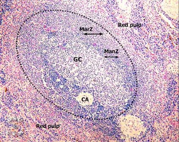
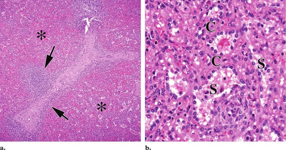
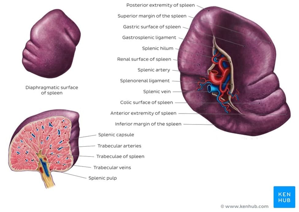
The spleen is a fist sized organ located in the left upper quadrant of the abdomen. It is the largest lymphoid organ and thus the largest filter of blood in the human body. The spleen has a unique location, embryological development and histological structure that differs significantly from other lymphoid organs. <br />Special histological features define several important functions of the spleen, such as filtering blood, maintaining immune response balance and recycling iron. The spleen can also serve as a reservoir for additional blood in situations of acute or chronic blood loss (such as bleeding or anemia), as well as an alternative site for hematopoiesis (formation of blood cells and platelets) outside of bone marrow. Even though the spleen has a few unique functions that can't be replaced by other lymphoid organs, it is not a vital organ and people can live without it. <br />Structure<br /> Being an intra-peritoneal organ, the spleen is covered by a layer of visceral peritoneum. Underneath the peritoneum is the capsule of the spleen, encasing its parenchyma.<br />The capsule of the spleen consists of dense irregular fibroelastic tissue. The connective tissue of the capsule contains contractile cells called myofibroblasts. By producing weak contraction of the capsule, these cells help to discharge the blood stored within the spleen into the circulation. The capsule also allows the spleen to significantly increase in size when necessary and discharge a large amount of blood to contribute to the tissues oxygenation, like during physical exercise. At the level of the hilum, the capsule splits into several septae called trabeculae which penetrate into the parenchyma of the spleen and partly divide its tissue.<br /> <br /> Spleen diaphragmatic surface <br />Like every other organ, the spleen consists of stroma and parenchyma. The stroma of the spleen is composed mainly of a network of reticular connective tissue. This mesh provides support for blood cells and cells of the immune system (lymphocytes, macrophages, and dendritic cells). The parenchyma of the spleen is divided into two functionally and morphologically distinct compartments (red pulp and white pulp) divided by a tissue layer called the marginal zone. Outside the marginal zone is the perifollicular zone which contains sheathed capillaries and blood-filled spaces without endothelial lining. <br /><br /><br />The red pulp <br />The red pulp occupies the majority of the stromal tissue of the spleen. It consists of the cords of Billroth and splenic sinusoids. The cords of Billroth (splenic cords) are the cellular aggregations supported by the reticular connective tissue. They appear as stripes and consist of of macrophages, plasmocytes and blood cells.<br />Splenic sinusoids are found between the cords of Billroth. They are filled with blood and give the red pulp its distinguishable red appearance. Blood slowly flows through the sinusoids where it is exposed to macrophages from the cords of Billroth, patiently waiting for foreign antigens that can appear in the blood. In a nutshell, the red pulp functions as a blood filter for various toxins, destroying them before they enter systemic circulation and get the chance to spread throughout the body and damage other organs.<br /><br /> <br />Normal splenic red pulp shows normal red pulp ( * ) shows splenic sinuses (S) The splenic cords (C) <br />The white pulp<br />The white pulp of the spleen is made of three different compartments: Periarterial lymphoid sheath (PALS), lymphoid follicles and the marginal zone.<br />The PALS consists of a central artery (a branch of the splenic artery) surrounded by a sheath of lymphoid tissue. Here, the lymphoid tissue organized into two layers: The inner layer and outer layer. The inner layer is mainly composed of T lymphocytes which is why it is also called the T-zone. The outer layer has a more diverse cellular morphology, containing T and B lymphocytes.<br />The branches of central arterioles are surrounded by the sharply defined areas of B lymphocytes, comprising the lymphoid follicles of the spleen. There are two types of lymphoid follicles depending on the features of the B lymphocytes that comprise them: Primary follicles and secondary nodules.<br />A follicle that consists mainly of small, immature lymphocytes is called a primary follicle. However, most nodules found in the spleen are secondary nodules that arise from primary follicles as the lymphocytes mature and increase in size. They differ from primary follicles by featuring a distinctive centrally positioned zone called the germinal center. The germinal centers are the sites where lymphocytes mature and acquire the ability to produce antibodies. So, seeing the germinal center is a sign that lymphoid tissue is responding to an antigen. Other than B lymphocytes, the germinal centers also contain follicular dendritic cells (FDC) which also increase in number after antigen activation. They support B lymphocytes, initiate and modulate their immune response.<br />The marginal zone is found on the very edge of the lymphoid follicles, containing different immune cells that are well equipped for starting an appropriate immune response.<br />