
Role of Contrast Enhanced Cardiac Magnetic Resonance Imaging in Assessment of Ischemic Heart Disease م.م حسين عايد حسين
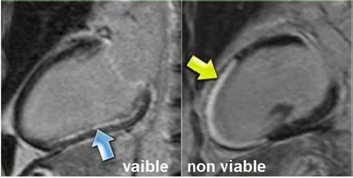
Role of Contrast Enhanced Cardiac Magnetic Resonance Imaging in Assessment of Ischemic Heart Disease<br />Abstract<br />Recently cardiac magnetic resonance imaging has emerged as a prime player in the clinical and preclinical detection of ischemic heart disease. Delayed-enhancement MR imaging is an extremely accurate method in which it can identify the presence, location, and the size of myocardial fibrosis (dense myocardial fibrosis) as a result of ischemic heart disease.<br />After delayed contrast enhancement nonviable (dead) myocardial tissues appear with increased signal intensity. The study of myocardial viability is of great importance in the orientation and management of patients requiring myocardial revascularization. The technique of delayed enhancement is accurate and has transformed the study of viability into an easy test, not only for the detection of fibrosis but also as a binary test, detecting whether it’s viable or not. concerning the delayed contrast enhancement, fibrosis is greater than 50% of the segmental area which is considered as non-viable, whereas fibrosis which is equal or below 50%, it is considered viable. <br /> <br />Figure (1) Blue Arrow Refers to Subendocardial Infarction with Scar Transmurality Less than 50% where Consider Viable, while Yellow Arrow Refers to Transmural Infarction with Transmurality More than 50% where Consider Non-Viable.