
An article titled "What is an MRI and where is it used" by Engineer Muhammad Abdul Karim Razouki
What is an MRI and where is it used?
Magnetic resonance imaging (MRI) is a medical imaging technique that uses a magnetic field and computer-generated radio waves to create detailed images of the organs and tissues in your body.
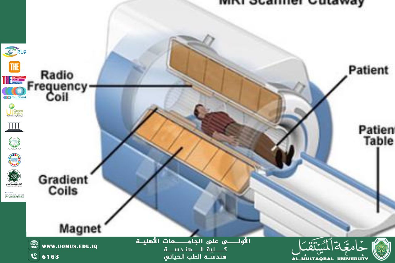
Most MRI machines are large, tube-shaped magnets. When you lie inside an MRImachine, the magnetic field inside works with radio waves and hydrogen atoms in your body to create cross-sectional images — like slices in a loaf of bread.
Why it's done
MRI is a noninvasive way for a medical professional to examine your organs, tissues and skeletal system. It produces high-resolution images of the inside of the body that help diagnose a variety of conditions.
MRI of the brain and spinal cord
MRI is the most frequently used imaging test of the brain and spinal cord. It's often performed to help diagnose:
Aneurysms of cerebral vessels.
Conditions of the eye and inner ear.
Multiple sclerosis.
Spinal cord conditions.
Stroke.
Tumors.
Brain injury from trauma.
MRI of the heart and blood vessels
MRI that focuses on the heart or blood vessels can check:
Size and function of the heart's chambers.
Thickness and movement of the walls of the heart.
Extent of damage caused by heart attacks or heart disease.
Structural problems in the aorta, such as aneurysms or dissections.
Inflammation or blockages in the blood vessels.
MRI of other internal organs
MRI can check for tumors or other irregularities in many organs in the body, including the following:
Liver and bile ducts.
Kidneys.
Spleen.
Pancreas.
Uterus.
Ovaries.
Prostate.
MRI of bones and joints
MRI can help look for:
Joint issues caused by traumatic or repetitive injuries, such as torn cartilage or ligaments.
Disk problems in the spine.
Bone infections.
Tumors of the bones and soft tissues.