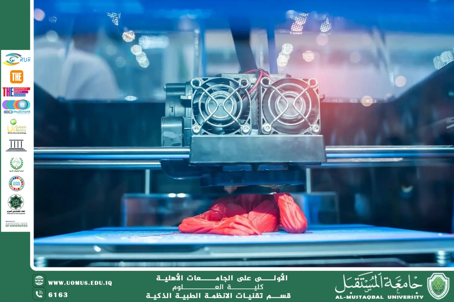
Printed Tissue Technology: How 3D Printing Contributes to the Production of Human Tissues (Asst. Lect. Ali Salim Halim)
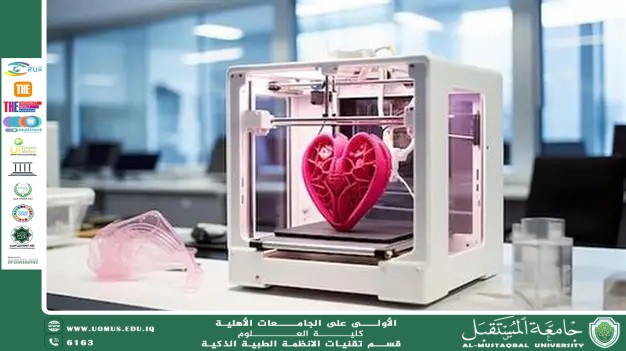
Printed tissue technology is considered one of the most prominent applications of three-dimensional (3D) printing in the medical field, representing a qualitative leap in tissue engineering and regenerative medicine. This technology enables the fabrication of biological structures that closely mimic human tissues in both structure and function. It relies on what is known as bioprinting, which applies the principles of Tissue Engineering to integrate living cells with supportive biomaterials known as “bioinks,” in order to construct three-dimensional tissues capable of growth and development within a suitable biological environment.
3D printing contributes to the production of human tissues by transforming medical images obtained from technologies such as magnetic resonance imaging (MRI) and computed tomography (CT) scans into highly accurate digital models representing the required organ or tissue. Specialized bioprinters then deposit successive layers of living cells and biomaterials according to a predefined design that takes into account spatial cell distribution, vascularization, and the mechanical properties of the tissue. This layered precision allows for the creation of complex structures that closely resemble the microscopic architecture of natural tissues, thereby enhancing their ability to integrate into the human body and perform vital biological functions.
One of the key advantages of printed tissues is the possibility of personalization using cells derived from the patient. This significantly reduces the likelihood of immune rejection after transplantation and minimizes the long-term need for immunosuppressive drugs. Additionally, this technology accelerates pharmaceutical research by enabling the production of laboratory-based human tissue models used to test drug efficacy and toxicity before progressing to clinical trials. This approach improves the accuracy of results and reduces reliance on animal models.
In the field of organ transplantation surgery, printed tissues offer promising solutions to the global shortage of donor organs. Bioprinting can be used to produce tissue patches or functional components of organs such as skin, cartilage, and certain parts of the liver or kidney, aiming to repair damaged tissues or support diseased organs without requiring complete replacement. Ongoing research also focuses on developing fully printed organs that incorporate intricate vascular networks capable of sustaining cellular life after transplantation, a major scientific challenge that remains at the forefront of current studies.
At the surgical level, printed tissues enable preoperative planning through biologically accurate models that replicate a patient’s anatomy, assisting surgeons in simulating complex procedures and reducing both operative time and associated risks. They can also be applied in reconstructive procedures following tumor removal or severe injuries, thereby improving both functional and aesthetic outcomes for patients.
Despite significant progress, technical and ethical challenges remain, including ensuring adequate vascularization within printed tissues, achieving long-term stability after transplantation, and establishing appropriate regulatory and legal frameworks for clinical application. Nevertheless, with continuous advancements in molecular biology, biomaterials science, and biomedical engineering, printed tissue technology is expected to become a fundamental pillar in the future of regenerative medicine and organ transplantation surgery, contributing to improved quality of life and reduced mortality rates associated with organ failure.
Al-Mustaqbal University – The First University in Iraq.