Computed Tomography (CT-Scan)
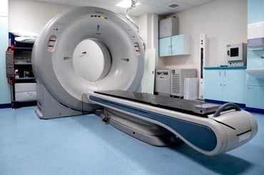
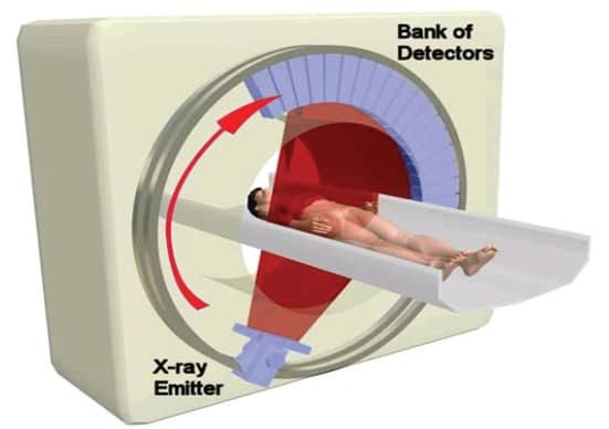
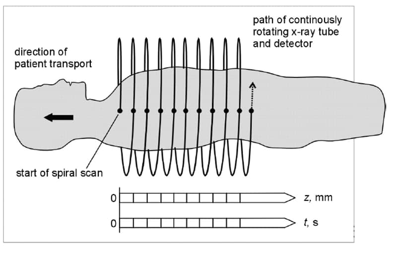
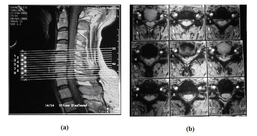
A CT-Scan is a radiological imaging test that creates detailed images of the body according to Medline pulse. It may be used to diagnose an infection, guide a surgeon to the correct area throughout biopsy, identify masses tumor within the body, or examine blood vessels. This imaging machine rotates around the patients while shooting X-Ray beams towards them. A computer that is connected to the machine creates separated slices or images of the body.<br />In the period between 1957 and 1963, A. Cormic independently created a method of determining the radiation absorption in human body. He found that there is a small difference in radiation absorption through different tissues inside the human body. William Oldendorf and Godfrey Hounsfield in the early of 1970’s created and developed a computerized tomography (CT-Scan). Their test produced a slice image which required further improvements and it cannot be used yet. This test uses a single ray beam which rotates around the patient. The process takes very long time to complete one test. To reduce the time required to complete a single CT-Scan test several improvements were made. The CT-scan improved to have an X-Ray source that produces fan beam. This new device rotates in 360o around a central axis (patient). The density of material is found by solving the complex image algorithm. The quality of the image is affected by patient axis movements. In 1980’s, a CT-Scan was developed to create a single slice image in less than 1 second. In 1990’s, the spiral CT-Scan was firstly introduced, and the scan time for a single slice image was reduced to 0.5 second. The slice can be imaged for brain, skull, spin cord, and bones. The slice is two dimensional, and the quality of the images provides the capability to distinguish between black, gray, and white area.<br />