Anesthesia for diagnostic imaging
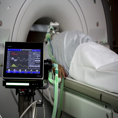
Anesthetists are increasingly working within medical imaging departments. However, the environment remains potentially hazardous, and the equipment unfamiliar. Ensure that experienced, trained assistance and full monitoring are available. Familiarizes yourself with the staff and surroundings. Locate the nearest resuscitation facilities (self- inflating bag/ mask, portable<br />O2, ‘crash’ trolley and defibrillator) and confirm that your assistant and the radiographers also know where these are located.<br /><br />Indications for an aesthesia<br />• Young or uncooperative children. Infants (under 2mo) may sleep through a scan if given a feed and wrapped up well.<br />• Older children or adults with psychological, behavioral or movement<br />Disorders.<br />• Acute trauma patients or patients receiving intensive care.<br />• Interventional procedures under ultrasound, CT or MRI guidance that<br />require analgesia, sedation or anesthesia.<br />Anesthesia: general points<br />Patients requiring an aesthesia for elective scans have a range of problems. Check the indications for the scan and the nature of the underlying pathology, e.g. developmental delay, epilepsy, malignancy, psychiatric disease or movement disorders. Beware the ‘undiagnosed’ pediatric patient and syndromes with CVS manifestations.<br />• Choice of sedation or GA depend upon individual patient needs, the nature of the scan and the skills of the anaesthetist. Check whether the anesthetic machines are using piped gases or cylinders. If using cylinders, confirm that a full spare O2 cylinder is<br />Immediately available.<br />• Plan the location of the anesthetic machine, suction, monitoring and the configuration and routing of the breathing system with<br />Radiographers in advance. Ensure the breathing circuit is sufficiently long for any gantry movement.<br />• Decide where to induce the patient— a dedicated induction area may not be available or may be very small. It is usual to induce on a tilting trolley, then transfer to the scanner when anaesthetized.<br />• Certain equipment configurations (e.g. anesthetic machine in the scan room and monitors in the control room) may require two anesthetists to manage the patient safely.<br />• Satisfactory recovery facilities must be available, i.e. an appropriately equipped recovery bay staffed by an experienced recovery nurse near the scanner. If not available, arrangements for safe transfer of the patient to an operating department recovery room must exist.<br />• Intensive care patients requiring diagnostic imaging should be managed with full transport monitoring and ventilator support. Ideally, the ICU medical team should supervise the patient and review the scan with the reporting radiologist before return to the ICU.<br />Anesthesia for CT<br />• CT scanning does not restrict the type of equipment used, but space can be limited, so compact machines and monitors are ideal.<br />• The patient, anesthetic machine and monitors must all be visible from the control room.<br />• The patient’s head is usually accessible during CT scanning, so an SGA may be used if airway protection is not required.<br />• Anesthesia or sedation sufficient to produce immobility and lack of<br />Awareness is all that is required for diagnostic procedures.<br />Hazards<br />• CT scanning uses ionizing radiation, so it is preferable for the anaesthetist to monitor the patient from outside the scan room. If it<br />Is necessary to remain near the patient, wear appropriate radiation protection and use barriers if available.<br />• Cannula, catheters, drains and ETTs can pull out during movement of the patient through the scanner— check nothing snags beforehand.<br />Contrast media<br />• IV contrast media for X- ray imaging are usually iodine- based, non- ionic, water- soluble compounds. Agents may trigger allergic reactions (ask about iodine sensitivity).<br />• Radiographers will usually give IV contrast, but you may be asked to administer it in anaesthetized/ pediatric patients. The radiographers should ensure the correct volume (dependent on preparation, investigation, age and weight) is provided according to local policy.<br />• Some ‘dynamic’ investigations (e.g. aortography) require contrast to be administered while the scan is occurring.<br />• Contrast is viscous and can be difficult to inject through small cannula<br />Or injection ports.<br />• Automated contrast injectors should not be connected to standard central venous lines. The high pressure developed by the rapid injection of viscous medium down a long, narrow lumen can burst the line. Power Line® catheters allow power- injection of contrast media.<br />• Contrast media may cause kidney injury in patients with dehydration or impaired renal function, so ensure patients are adequately hydrated. Lactic acidosis can be precipitated in patients taking iguanids<br />(Metformin)— Ideally avoid for 48h before and after the scan.<br />Practical considerations<br />• Move metal- containing objects (e.g. ECG leads, pressure transducer cables) away from the area being scanned to prevent X- ray artefact.<br />• Thoracic and abdominal scans may require ‘breath- holds’ of a few seconds to reduce respiratory movement artefact. Both paralyzed and spontaneously breathing patients can be ventilated manually, and their lungs held in inspiration for each individual scan.<br />• The patient’s arms ideally need to be positioned above the head during thoracic and abdominal scans. Soft Velcro straps attached to the gantry or wide adhesive tape are useful for securing the limbs.<br /><br />Miaad adnan abdul salam<br />Iraqi board of anesthesia and intensive care