The Role of IVIM diffusion in breast cancer diagnosis BY DR MOHANED AHMED SAHIB
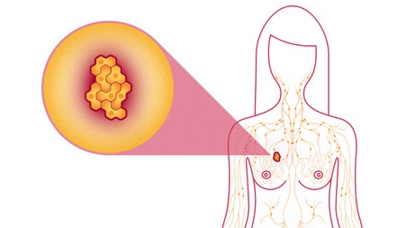
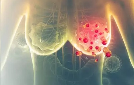
Intravoxel Incoherent Motion (IVIM) diffusion-weighted imaging (DWI) has emerged as a promising technique in the diagnosis and characterization of breast cancer. This article provides a comprehensive overview of the role of IVIM-DWI in distinguishing malignant from benign breast lesions, highlighting its diagnostic performance, methodological approaches, and clinical applications. We discuss the IVIM parameters, including the true diffusion coefficient (D), pseudodiffusion coefficient (D*), and perfusion fraction (f), and their utility in enhancing diagnostic accuracy. The study synthesizes findings from recent research, including meta-analyses and clinical studies, to evaluate the effectiveness of IVIM-DWI in breast cancer diagnosis and its potential to complement traditional imaging techniques.<br /><br />Introduction<br />Breast cancer is one of the most common cancers in women worldwide, and accurate diagnosis is crucial for effective treatment and patient outcomes. Magnetic Resonance Imaging (MRI) has become a vital tool in breast cancer diagnosis due to its high sensitivity and specificity. Within the realm of MRI, Diffusion-Weighted Imaging (DWI) and specifically Intravoxel Incoherent Motion (IVIM) DWI have gained significant attention for their ability to characterize tissue microstructure and detect subtle changes associated with cancer.<br /><br />IVIM-DWI extends the traditional DWI by accounting for both the diffusion of water molecules and the perfusion effects within the tissue, providing a more comprehensive understanding of tissue characteristics. This technique has shown promise in differentiating between malignant and benign breast lesions, as well as in characterizing the histological and molecular subtypes of breast cancer.<br /><br />Method<br /><br />Data Collection<br /><br />This study involved a systematic review of literature on the use of IVIM-DWI in breast cancer diagnosis. Databases such as PubMed, Embase, and Web of Science were searched for studies published over the last decade. The search included keywords related to IVIM-DWI, breast cancer diagnosis, and comparative studies with other imaging techniques.<br /><br />Study Selection<br /><br />Studies were selected based on their focus on the diagnostic performance of IVIM-DWI in differentiating malignant and benign breast lesions. Only studies that provided detailed information on IVIM parameters, such as D, D*, and f, were included. Studies with small sample sizes or those lacking robust statistical analysis were excluded.<br /><br /><br /><br /><br /><br />IVIM Parameters<br /><br />IVIM-DWI involves the calculation of several key parameters:<br /><br />- **True Diffusion Coefficient (D):** Represents the diffusion of water molecules in the tissue.<br />- **Pseudodiffusion Coefficient (D*):** Reflects the perfusion-related diffusion motion in the microcirculation.<br />- **Perfusion Fraction (f):** Indicates the volume ratio between the microcirculation perfusion effect and the total diffusion effect in a voxel[2][3][5].<br /><br />These parameters are calculated using the IVIM model, which can be simplified to reduce the complexity of data acquisition and analysis.<br /><br />Simplified IVIM Approach<br />Recent studies have explored the use of simplified IVIM approaches, which involve acquiring DWI sequences with fewer b-values (e.g., 0, 50, 250, 800 s/mm^2). This method simplifies the calculation of IVIM parameters without the need for complex fitting procedures, making it more practical for clinical applications[1][2].<br /><br />Statistical Analysis<br />The diagnostic performance of IVIM-DWI was evaluated using metrics such as sensitivity, specificity, and the area under the curve (AUC) of the receiver operating characteristic (ROC). Meta-analyses were conducted to pool the results from multiple studies, assessing the standardized mean difference (SMD) and 95% confidence intervals of the IVIM parameters[2].<br /><br />Results<br />Diagnostic Accuracy<br /><br />The studies reviewed demonstrated that IVIM-DWI parameters are superior to traditional apparent diffusion coefficient (ADC) values in differentiating malignant from benign breast lesions.<br /><br />- **Sensitivity and Specificity:** The D value showed the best diagnostic performance with a sensitivity of 86%, specificity of 86%, and an AUC of 0.91. ADC values also performed well, with a sensitivity of 76%, specificity of 79%, and an AUC of 0.85[2].<br />- **IVIM Parameters:** The combination of D and f values further improved diagnostic accuracy, achieving a high diagnostic accuracy of 93.7% in one study[1].<br />- **Clinical Applications:** IVIM-DWI was effective in distinguishing invasive ductal carcinoma from ductal carcinoma in situ (DCIS) and in identifying lymph node metastasis, histologic grade, and hormone receptor status[2].<br /><br />Clinical Studies<br />Several clinical studies have validated the use of IVIM-DWI in breast cancer diagnosis:<br /><br />- **Breast Lesion Differentiation:** IVIM-DWI was used to differentiate between malignant and benign breast lesions with high accuracy. The technique was particularly useful in cases where diffusion and perfusion effects oppositely influence the DWI signal decay[1][2][5].<br />- **Tumor Heterogeneity:** Histogram analysis of IVIM parameters based on tumor heterogeneity helped in predicting histopathological types and molecular subtypes of breast tumors[4].<br /><br />Comparative Studies<br /><br />Comparative studies with other imaging techniques, such as dynamic contrast-enhanced MRI (DCE-MRI), showed that IVIM-DWI can serve as a valuable adjunct to improve diagnostic accuracy:<br /><br />- **Combination with DCE-MRI:** The use of IVIM-DWI in addition to DCE-MRI enhanced the accuracy of differentiating benign and malignant lesions, especially in cases with suspicious contrast enhancement[1][3].<br /><br /> <br />Discussion<br /><br />The integration of IVIM-DWI into breast cancer diagnosis offers several key advantages:<br /><br />Enhanced Diagnostic Precision<br /><br />IVIM-DWI provides a more nuanced understanding of tissue characteristics by separating the effects of diffusion and perfusion, which is particularly useful in distinguishing between malignant and benign lesions. The D value, which represents true diffusion, has been shown to have the best diagnostic performance among IVIM parameters[2].<br /><br />Clinical Utility<br /><br />IVIM-DWI can be used as a stand-alone tool or as an adjunct to other imaging techniques like DCE-MRI. Its ability to characterize tumor microstructure and detect subtle changes makes it valuable for early disease detection, treatment monitoring, and predicting responses to neoadjuvant therapy[1][2][5].<br /><br />Practical Considerations<br /><br />The simplified IVIM approach, which requires fewer b-values, makes the technique more feasible for clinical use. However, standardization of IVIM protocols and quality control measures are essential to ensure consistent results across different studies and clinical settings[1][2].<br /><br />Challenges and Limitations<br /><br />Despite its benefits, IVIM-DWI faces some challenges:<br />- **Technical Variability:** Differences in b-value selection, acquisition protocols, and analysis methods can affect the consistency of IVIM parameters across studies.<br />- **Clinical Integration:** Resistance to change and the need for training among healthcare professionals can hinder the adoption of IVIM-DWI in routine clinical practice.<br />- **Sample Size and Heterogeneity:** Many studies have small sample sizes, and the heterogeneity in patient populations and lesion types can impact the generalizability of the results[2].<br /><br />Conclusion<br />Intravoxel Incoherent Motion diffusion-weighted imaging has emerged as a valuable tool in the diagnosis and characterization of breast cancer. The technique offers enhanced diagnostic precision by distinguishing between the effects of diffusion and perfusion, making it superior to traditional ADC values in differentiating malignant from benign breast lesions. The simplified IVIM approach and its ability to complement other imaging techniques, such as DCE-MRI, further enhance its clinical utility.<br /><br />While there are challenges to be addressed, including standardization of protocols and clinical integration, the evidence supports the potential of IVIM-DWI to improve diagnostic accuracy and patient outcomes in breast cancer. Continued research and clinical validation are necessary to fully realize the benefits of this technique in routine clinical practice.<br />