

X-Rays Imaging in Medicine
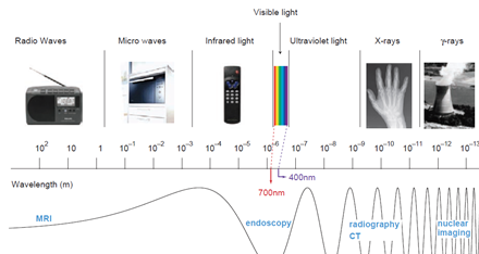
Introduction to X-ray and CT –Scan<br /><br />• X-rays were discovered by Wilhelm Konrad Rontgen in 1895 while he was experimenting with cathode tubes.<br />• Rontgen found out that x-ray was attenuated in a different way by various kinds of materials and that it could, like light, be captured on a photographic plate . <br />• This opened up the way for its use in medicine.<br />• The first “Rontgen picture” of a hand was made soon after the discovery of X-rays.<br />X-rays:<br /> X-rays : are electromagnetic waves . The wavelength for X-rays is on the order of Angstroms (10−10 m) The nature of X-rays as short-wave electromagnetic radiation was established in 1912. Electromagnetic radiation consists of photons . X- RAY GENERATION:<br />• X-rays are generated in an X-ray tube, which consists of a vacuum tube with a cathode (tugestone fillament ) and an anode (tungestone target) The x-ray beam is produced by bombarding a tungsten target with an electron beam within an x-ray tube <br />X-ray detectors:<br />The detector s can be :<br /> 1- A screen – film combination : in which a film is sandwiched between two screens , Screen–film detector ( Radiography ) The film contains an emulsion with silver halide crystals (e.g., AgBr) When exposed to light, the silver halide grains absorb optical energy and undergo a complex physical change . <br /> 2 - An image intensifier coupled to a camera ( Fluoroscopy ),<br /> 3 - A cassette containing a storage phosphor plate ( computed radiography ) <br />4 - An active matrix flat panel detector or dual-layer detector (digital radiography ) <br />Interaction with matter:<br />When x-ray beam pass through a matter they will interact in the following ways : <br />• Trasmited : pass through unaffected . A primary or direct radiation . <br />• Absorbed : transfering to the matter all of their energy (the photon disappearing completely ) <br />• Scattered : diverted in a new direction , with or without loss of energy and so leave the material as scattered or secondary radiation . <br />The X-ray image is formed by the transmitted photons. those that are absorbed or scattered represent attenuation by matter . <br />An understanding of how the properties of X-ray and the materials through which they travel affect the relative amount of attenuation and transmission gives an understanding of how the X-ray image is formed. <br />The interactions with tissues , depend on <br />- the energy of the photon , E = hxf , f = c/Y . In most radiological examinations the voltage used is typically in the range from 50 to 125 kV. <br />- the atomic number of the interacting matter ,the higher the atomic number the more the attenuation , so the contrast media used in radiography to opacify certain part of the body should have high atomic number .<br />The most important interactions are the following : <br />• photoelectric absorption. A photon can be absorbed by an atom while its energy excites an electron.<br />• Compton scattering. A second possibility is that the photon transfers only part of its energy to eject an electron with a certain kinetic energy ,the electron then escapes in another direction.<br />According to x- ray attenuation in the tissues ( x – ray penetration) ,the radiographic appearace can be graded into : <br />• Tranceradiant as gases <br />• Radiolucent or trancelucent as in fatty tissue <br />• Mild radio radio-opague as fluid , muscle ..<br />• Moderate radio-opague as bones and calcifications <br />• Dense radio-opague as metals and contrasts<br />Clinical use (DIAGNOSTIC):<br />Radiography ( static ) : Conventional and digital ( CR and DR )<br />Fluoroscopy ( dynamic ) Analogue or digital : Used in contrast studies and interventional procedures <br />CT scan (Computed tomography scan )<br />Computed tomography or CT is an imaging modality that produces cross-sectional images representing the X-ray attenuation properties of <br />the body.<br />Types and generations :<br />A- CAT (computed axial tomorgaphy ) scan<br />Single-slice CT , Circular CT<br />The most straight forward way to image an entire volume is to scan a number of consecutive slices by circular tube–detector rotations alternated with small table shifts.<br />B- Spiral CT (Helical CT) <br /> A technique that is widely used nowadays is helical CT. The X-ray tube rotates continuously around thepatient, just as in 2D CT. At the same time, the patientis slowly translated through the gantry.<br /><br />Multidetector spiral CT scan ( 16 slice , 32 slice , 64 ,128 and 256 slice) . <br />In modern CT scanners, the detector array consists of multiple detector rows, in order to measure several slices per rotation of the X-ray tube.<br /><br /><br /><br />