A scientific article by the lecturer, Asst. Lecturer Hassan Qahtan Kazem Al-Hussein (Optic Nerve Damage: Causes, Symptoms, Diagnosis, and Treatment)
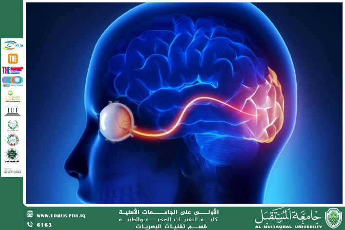
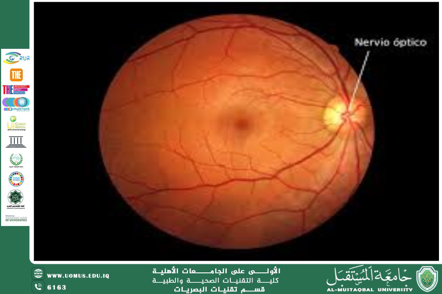
🟦 Introduction<br />The optic nerve is a crucial component of the visual system, responsible for transmitting visual information from the retina to the brain. Optic nerve damage (Optic Neuropathy) is a serious condition that can lead to visual impairment or permanent vision loss if not diagnosed and managed early.<br />This article aims to highlight the common causes of optic nerve damage, provide a clinical framework for diagnosis and follow-up, and review the latest available therapeutic approaches.<br />🟦 I. Anatomy and Function of the Optic Nerve<br />The optic nerve (cranial nerve II) consists of approximately one million nerve fibers originating from retinal ganglion cells. It transmits electrical impulses to the visual cortex in the occipital lobe of the brain.<br />The anatomical pathway includes:<br />• Optic disc<br />• Optic canal<br />• Optic chiasm<br />🟦 II. Main Causes of Optic Nerve Damage<br />1. Glaucoma:The most common global cause. Chronic elevated intraocular pressure leads to progressive optic nerve fiber damage.<br />2. Optic Neuritis:Often associated with Multiple Sclerosis (MS). Affects primarily young adults, especially women.<br />3. Ischemic Optic Neuropathy:Due to impaired blood flow, especially in patients with diabetes and hypertension.<br />4. Tumors:Orbital or intracranial masses may compress the optic pathway.<br />5. Toxins and Medications:Exposure to substances like methanol or excessive use of drugs such as Ethambutol.<br />6. Trauma:Direct or indirect mechanical injury to the optic nerve.<br />🟦 III. Clinical Symptoms of Optic Nerve Damage<br />• Gradual or sudden decrease in visual acuity<br />• Color vision distortion (Dyschromatopsia)<br />• Blurred vision or central blind spot (Central Scotoma)<br />• Visual field loss<br />• Ocular pain during eye movement (particularly in optic neuritis)<br />🟦 IV. Diagnostic Approach<br />1. Clinical Examination:<br />o Visual acuity and field testing<br />o Pupillary light reflex (Relative Afferent Pupillary Defect - RAPD)<br />o Color vision testing (e.g., Ishihara test)<br />2. Fundoscopy:<br />o Optic disc pallor or atrophy<br />o Disc swelling or blurred margins (in cases of inflammation)<br />3. Optical Coherence Tomography (OCT):<br />o Measures Retinal Nerve Fiber Layer (RNFL) thickness and optic disc morphology.<br />4. Magnetic Resonance Imaging (MRI):<br />o To assess central nervous system causes, such as tumors or demyelination.<br />5. Visual Evoked Potential (VEP):<br />o Evaluates electrical signal transmission from the eye to the brain.<br />🟦 V. Treatment Options<br />1. For Glaucoma:<br />o Intraocular pressure-lowering drops (e.g., prostaglandin analogs)<br />o Laser therapy or surgery when pharmacologic treatment fails<br />2. For Optic Neuritis:<br />o High-dose intravenous corticosteroids<br />o Treatment of the underlying condition (e.g., MS)<br />3. For Ischemic Causes:<br />o Control of blood glucose and blood pressure<br />o Use of anticoagulants when indicated<br />4. For Toxic Etiologies:<br />o Immediate cessation of exposure<br />o Supportive and detoxification measures<br />5. Rehabilitative Measures:<br />o Low vision aids (LVAs)<br />o Visual training programs<br />o Referral to low vision centers for supportive care<br />🟦 Conclusion<br />Optic nerve damage presents a significant clinical challenge due to its diverse causes and the often irreversible nature of nerve injury.<br />Successful management depends on early detection, accurate assessment of the underlying pathology, and multidisciplinary collaboration involving optometrists, neurologists, and internal medicine specialists.<br />