A scientific article by the lecturer, Assist.Lect. Mohaimen Sameer Aref (Disorders of the Lacrimal System and Their Impact on Ocular Health and Visual Quality)
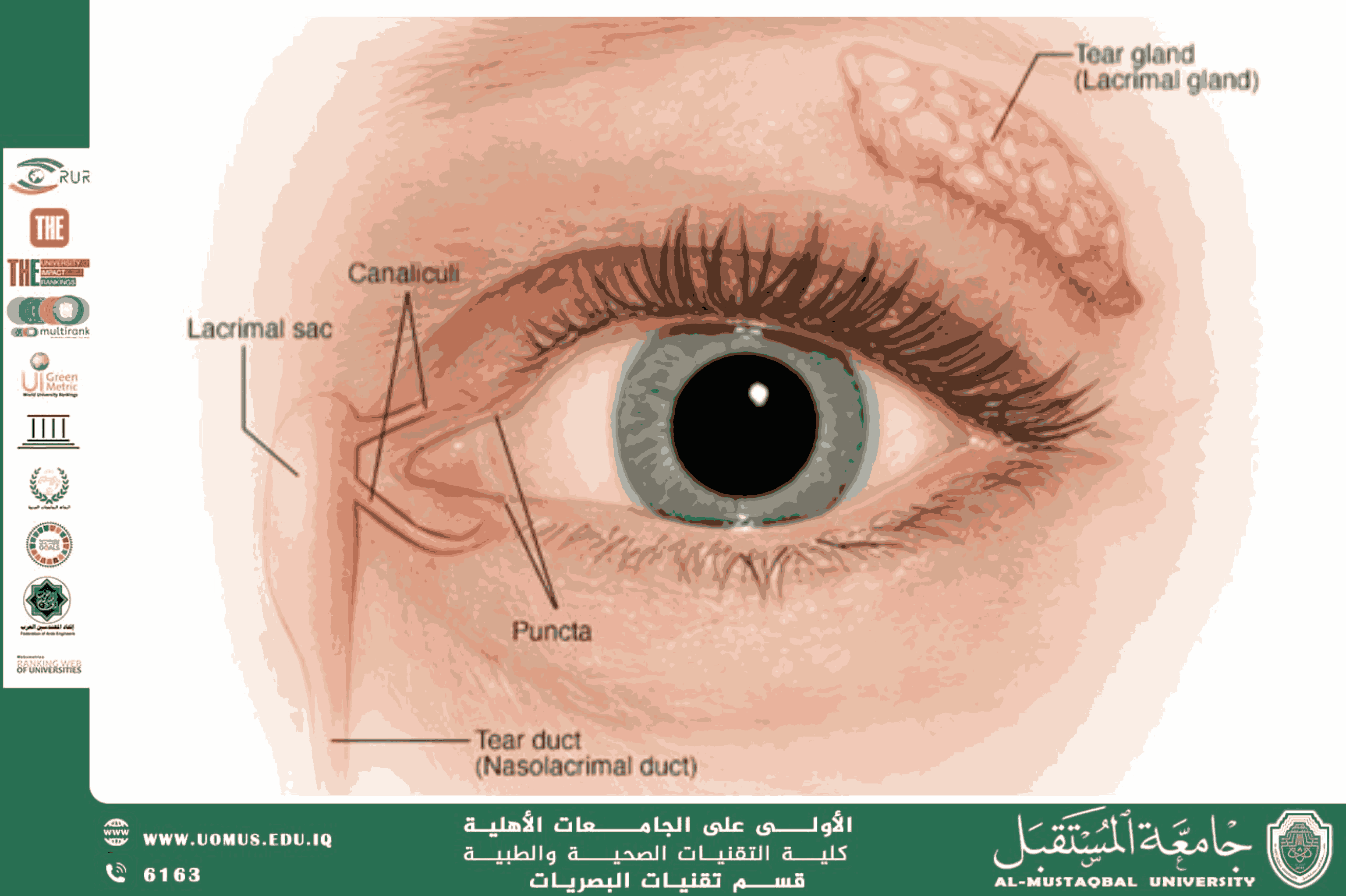
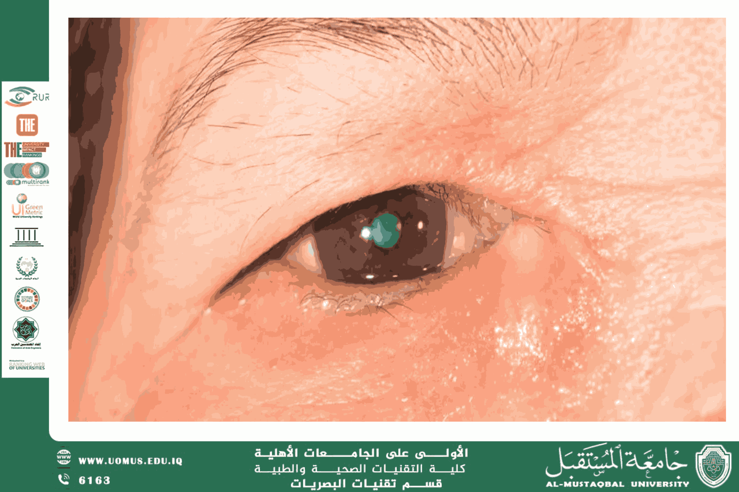
The lacrimal system is a vital component of the eye, playing an essential role in maintaining corneal and conjunctival hydration, providing a clear optical surface that enables efficient transmission of light, and protecting the eye against pathogens and foreign particles. Any dysfunction within this system may lead to a spectrum of disorders, ranging from mild dry eye disease to chronic infections and severe complications that can threaten corneal integrity and vision.<br />The lacrimal system consists of:<br />1.Main lacrimal gland and accessory glands: Responsible for producing the aqueous layer of the tear film.<br />2.Tear film: A trilaminar structure composed of a superficial lipid layer, an intermediate aqueous layer, and a basal mucin layer. Together, these ensure lubrication, ocular surface protection, and stabilization of the tear film.<br />3.Lacrimal drainage system: A network of puncta, canaliculi, lacrimal sac, and nasolacrimal duct that drains excess tears into the nasal cavity.<br />Major Lacrimal Disorders<br />1. Dry Eye Disease (DED)<br />This is the most prevalent lacrimal disorder, resulting from decreased tear production or excessive evaporation. Symptoms include burning sensation, foreign body sensation, and fluctuating visual clarity. DED is frequently associated with autoimmune disorders such as Sjögren’s syndrome, or with prolonged digital device use.<br />2. Dacryoadenitis<br />An inflammatory condition of the lacrimal gland caused by viral or bacterial infections, or systemic autoimmune diseases. It typically presents as painful swelling in the superotemporal region of the eyelid, sometimes accompanied by fever.<br />3. Nasolacrimal Duct Obstruction (NLDO)<br />Obstruction of the tear drainage pathway leads to epiphora (persistent tearing) and predisposes to recurrent infections. It is commonly observed in neonates due to congenital anomalies, but may also occur in adults as a result of chronic inflammation, trauma, or neoplasms.<br />4. Dacryocystitis<br />This is an infection of the lacrimal sac, often secondary to NLDO. It presents as a painful, erythematous swelling at the medial canthus, frequently with purulent discharge. Untreated cases may progress to abscess formation or orbital cellulitis.<br />Diagnostic Approaches<br />Diagnosis relies on medical history and clinical examination, supported by:<br />• Schirmer’s test: To assess aqueous tear secretion.<br />• Ocular surface staining: Using fluorescein or rose bengal dyes to evaluate tear film stability and corneal integrity.<br />• Imaging studies: Dacryocystography and computed tomography for structural assessment of the lacrimal drainage system.<br />Management Strategies<br />Treatment depends on the underlying disorder:<br />•Dry Eye Disease: Artificial tears, anti-inflammatory agents, and punctal occlusion in severe cases.<br />•Bacterial Infections: Topical or systemic antibiotics.<br />•Congenital NLDO in infants: Lacrimal sac massage, with probing or dacryocystorhinostomy (DCR) if obstruction persists.<br />•Adult NLDO: Often requires surgical intervention such as external or endoscopic DCR to re-establish tear drainage.<br />The lacrimal system is not merely a tear-producing mechanism but a crucial protective barrier that safeguards ocular clarity and visual function. Early recognition and appropriate management of lacrimal disorders are essential to prevent serious complications affecting the cornea and overall visual quality.<br />