Protocol for Routine MR Imaging of the Brain م.م علي هاني كريم
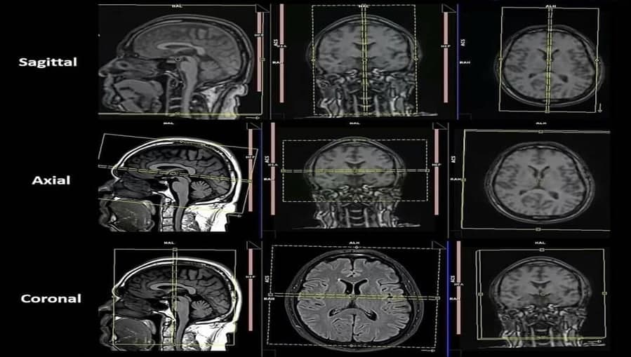
Protocol for Routine MR Imaging of the Brain <br />Ideally, imaging protocols for the brain should be as short as possible and focused on the clinical questions. Protocols should be standardized to ensure continuity over time. Because the diagnosis is based on experience, frequent changes in protocols should be avoided; moreover, frequent protocol changes may be confusing to the technologists operating the MRI equipment. Imaging protocols should be adapted to the equipment available. As a general rule, MR imaging studies of the brain should include at least two imaging planes and two ‘weightings’.the ‘traditional’ and ‘modern’ protocols for MR imaging of the brain. In the traditional screening protocol, the long TR sequence can be obtained with either a spin-echo (SE) or turbo spin-echo (TSE) technique. This sequence provides proton-density weighted images (PD-WI) and T2- W images (T2-WI). They are used to detect intraparenchymal signal abnormalities. Most pathological processes in the brain result in increased water content (vasogenic edema, cytotoxic edema, necrosis, or cyst formation) and are therefore readily identified on T2- WI. Small high-signal intensity (SI) lesions adjacent to the ventricles or subarachnoid spaces, e.g., periventricular white matter and cortical gray matter, may be missed on T2-WI because they cannot be differentiated from the cerebrospinal fluid (CSF), which is also hyperintense . These lesions are better appreciated on PD-WI, where the lesions are hyperintense but the SI from CSF is diminished .A more modern alternative is to use a T2- W sequence with dark CSF signal, such as fluid-attenuated inversion recovery (FLAIR) . In this T2- W sequence, the signal of CSF is attenuated by the use of a long inversion time, typically around 2000 ms. FLAIR provides excellent contrast resolution at brain CSF interfaces and improves the conspicuity of small white-matter lesions. Thus, in the modern imaging protocol, FLAIR and TSE T2-W sequences are used instead of the dual-echo PD-W and T2-W sequence (Table 4.2). The short repetition time (TR)/short echo time (TE) or T1-W sequence is used to evaluate the gross anatomy and structure of the brain. Moreover, T1-WI are often